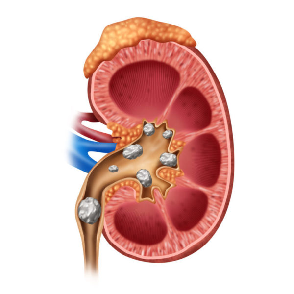
Kidney stones
Kidney stones (renal lithiasis, nephrolithiasis) are hard deposits made of minerals and salts that form inside kidneys.
Kidney stones form when the urine becomes concentrated or because of too much of a certain mineral in your urine, and the minerals to crystallize and stick together.
Kidney stones often don’t cause symptoms until they block urine drainage, which is generally when they have passed into the ureter. With obstruction, stones can be associated with severe flank pain, nausea, vomiting, blood in the urine, infection and kidney damage.
Passing kidney stones can be quite painful, but stones usually cause no permanent damage if they’re recognized and treated in a timely fashion. Management can range from medication to surgery.
When someone has a stone, some blood work and possibly urine work is done to identify factors which may predispose the patient to make stones.
Surgical Options:
Ureteroscopy: To remove a smaller stone in your ureter or kidney, your doctor may pass a thin lighted tube (ureteroscope) equipped with a camera through your urethra and bladder to your ureter.
Once the stone is located, special tools and lasers can snare the stone or break it into pieces that will pass in your urine. Your doctor may then place a small tube (stent) in the ureter to relieve swelling and promote healing. You will need anesthesia during this procedure, but it usually done in an outpatient setting.
Extracorporeal Shockwave Lithotripsy (ESWL): As long as stone is visible on KUB, your doctor can perform an outpatient procedure in which we use xray to identify the stone and focus ultrasonic shockwaves to break up the stone.
Percutaneous Nephrolithotomy (PCNL): This is an in-patient procedure generally indicated for stones greater than 2 cm in which access is obtained through the back, a large tract is dilated through the kidney to break up the stone.


